Courtesy: khanski clinical ophthalmology
Definition
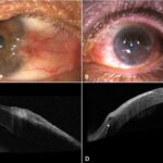
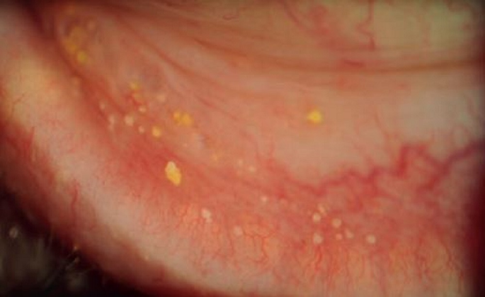
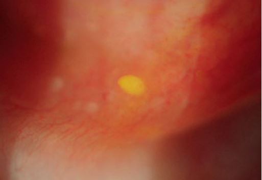
Conjunctival concretions are multiple tiny yellowish-whitish cysts or lesions which are deposits of epithelial debris. They are usually found subepithelially in the inferior tarsal and forniceal conjunctiva. They are commonly found in older people but can affect younger generations as well.
Causes of Conjunctival Secretions
The exact cause of conjunctival concretions is idiopathic but is known to result from degeneration of the conjunctiva. A common association is also found in chronic inflammation or conjunctivitis such as trachoma, vernal, severe atopic conjunctivitis and chronic meibomitis. According to a study by Haicl, conjunctival concretions were also found in patients with keratoconjunctivitis sicca (Dry Eye Syndrome). Also, some particular eye drops like sulfadiazine are known to have some links to conjunctival concretions when they recrystallize.
Pathology
Conjunctival Concretions are basically composed of protein-rich secretions (such as keratin) and degenerating epithelial cells of the conjunctiva. In inflammations, the aforementioned secretions and debris become trapped in subconjunctival pockets resulting in calcification. Sometimes large concretions may erode the overlying epithelium. Concretions of the conjunctiva are not so hard upon calcification as compared to other “lithiasis” that may occur at another part of the body like urinary tract.
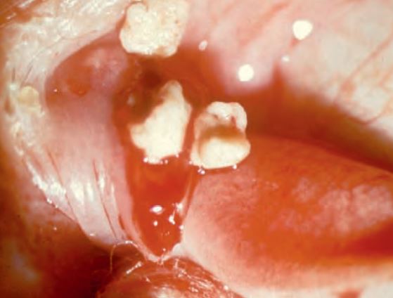
Sulfur Granules
Concretions resulting from canaliculitis (sulfur granules) are metabolic products of Actinomyces israelii which is responsible for most infections and other hydrogen sulfide-utilizing bacteria, and classically are expressed on canalicular compression. They are typically bigger in size than conjunctival concretions.
Courtesy: khanski clinical ophthalmology
Diagnosis
In most cases, the patient may not have any symptoms and as such wouldn’t present it as a complaint. An Optometrist or Ophthalmologist upon routine ocular examination using the slit lamp may notice about 1-2mm yellowish-white smallish lesion usually on the palpebral conjunctiva. These lesions can occur as singular, in multiple clusters, or in rare instances, they may merge together. They are predominantly situated on the lower tarsal and forniceal conjunctiva, although there are instances where they can be found in the superior conjunctiva as well. In situations where they are large enough, the degenerating epithelial cells may erode through the overlying epithelium resulting in local irritation and foreign body sensation.
Courtesy: khanski clinical ophthalmology
Management
In most instances, no treatment is required for conjunctival concretions since is it asymptomatic but in situations where there is marked irritation or foreign body sensation after eroding the overlying epithelium, then it may be removed. This can be done at the slit lamp with a needle or needlepoint forceps or a 30 gauge needle after the instillation of topical anaesthesia. After which a topical antibiotics may be given.