Ocular motility refers to the ability of the eyes to move in a coordinated manner in different directions and focus on objects at varying distances. This is a crucial aspect of vision that allows us to focus on objects and track them as they move. So how do our eyes move around without falling out? How is it able to follow fast-moving objects like the Ferrari cars without us feeling dizzy? To understand these we need to learn a brief anatomy of the eye.
Anatomy of the Eye
The eye is a complex organ that is responsible for translating light into electrical signals that the brain can interpret as images. The eye has several structures that work together to achieve this task. These structures include the cornea, iris, lens, retina, and optic nerve. The muscles in the eye are responsible for controlling its movement, allowing us to look in different directions.
The eye has six muscles that control its movement. These muscles known as ‘extraocular muscles’, work in pairs to move the eye up and down, side to side, and at an angle. The oculomotor nerve, trochlear nerve, and abducens nerve control the muscles that attach to the outer surface of the white part of the eye to the eye socket (orbit). Interestingly, the extraocular muscles have the densest ratio of motor neurons to muscle fibers of any muscle in the body, thus facilitating tremendously fine motor control and impeccable alignment of the eyes.
Types of Ocular Motility
There are three main types of eye movements: saccades, pursuits, and vestibulo-ocular reflex (VOR). Each type of eye movement plays a critical role in visual function.
Saccades
Saccades are rapid, jerky eye movements that allow us to quickly shift our gaze from one object to another. These movements are essential for scanning the environment and for reading. Saccades are controlled by the frontal eye fields and the superior colliculus in the brain.
During a saccade, the eye makes a rapid movement to the target, followed by a brief period of fixation. The movement is rapid and accurate, with precise control of the eye muscles.
Saccades can be affected by certain conditions, such as Parkinson’s disease and multiple sclerosis. In these conditions, saccades can become slower and less accurate, leading to difficulty with reading and other tasks.
Pursuits
Pursuits are slower, smooth, continuous eye movements that allow us to track moving objects. These movements are essential for activities such as watching a bird fly or following a moving vehicle. The movement is slower than a saccade and is more precise. Pursuits are controlled by the parietal eye fields in the brain.
Pursuits can be affected by certain conditions, such as cerebellar ataxia and Huntington’s disease. In these conditions, pursuits can become jerky and inaccurate, leading to difficulty with tracking moving objects.
Vestibulo-Ocular Reflex (VOR)
The vestibulo-ocular reflex (VOR) is a reflex that allows us to maintain visual stability while the head is moving. The VOR is essential for activities such as walking, running, and driving. The VOR is controlled by the vestibular system in the inner ear.
During a VOR, the eyes move in the opposite direction to the head movement, allowing us to maintain a stable visual image. The movement is automatic and does not require conscious effort.
The VOR can be affected by certain conditions, such as vestibular disorders and head injuries. In these conditions, the VOR can become impaired, leading to difficulty with maintaining visual stability during head movements.
Disorders of Ocular Motility
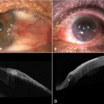
Several disorders can affect ocular motility, including strabismus, nystagmus, and amblyopia. These conditions can have a significant impact on a person’s quality of life and can affect their ability to perform daily activities.
Strabismus
Strabismus is a condition in which the eyes are misaligned, causing double or blurred vision. The condition can be caused by a muscle imbalance or a problem with the nerves that control the eye muscles. Strabismus can occur at any age, but it is most common in children. If left untreated, strabismus can lead to amblyopia.
To address refractive errors contributing to the condition, eyeglasses can be employed. Additionally, patching is utilized to strengthen the weaker eye and enhance binocular vision. Finally, surgery becomes an option to correct the muscle imbalance and straighten the eyes.
Nystagmus
Nystagmus is a condition in which the eyes make involuntary repetitive movements. The condition can be congenital or acquired and can be caused by a variety of underlying conditions, such as neurological disorders or drug toxicity. Nystagmus can cause problems with depth perception, poor visual acuity, and difficulty with reading.
Treatment options for nystagmus include eyeglasses, medication, or surgery. Eyeglasses can be used to correct refractive errors that may be contributing to the condition. Using large-print books, magnifying devices, and increased lighting can also be helpful. Though rare, surgery can be done to change the position of the muscles that move the eyes.
Amblyopia
Amblyopia, also known as lazy eye, is a condition in which one eye does not develop normal vision during childhood. A muscle imbalance, a refractive error, or a structural problem with the eye can cause this. Amblyopia can cause problems with depth perception, poor visual acuity, and difficulty with reading.
Treatment options for amblyopia include patching and vision therapy. Patching can be used to strengthen the weaker eye and improve binocular vision. Vision therapy can be used to improve eye teaming, focusing, and tracking.
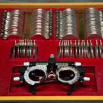
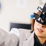
Evaluation of Ocular Motility
Several methods including the broad H test and Electrooculography can evaluate ocular motility.
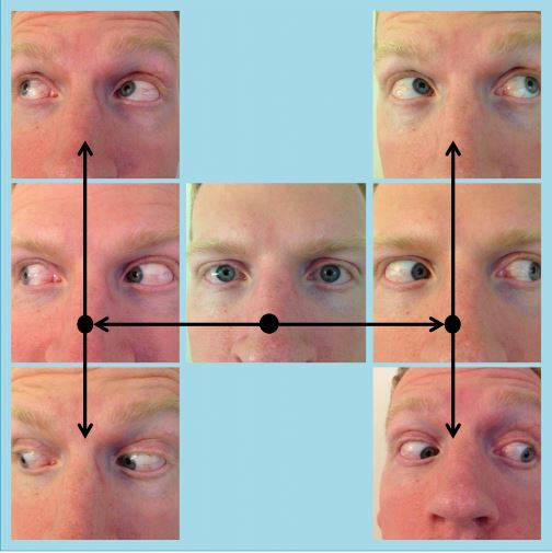
Broad H test
- Ask the patient to sit comfortably and look straight ahead.
- Move a target (such as a pen or a finger) in an H shape, starting from the center and moving from side to side. Move to the top left, top right, bottom left, and bottom right corners of the patient’s visual field.
- Observe the patient’s eye movements as they follow the target.
- Note any abnormalities in eye movements such as saccades, nystagmus, or strabismus.
- Repeat the test with the patient following the target with their head still and then with their head moving.
- Compare the results with normal eye movement patterns to determine if there are any abnormalities in the patient’s ocular motility.
Electrooculography
Electrooculography (EOG) is a non-invasive technique that evaluates ocular motility by measuring the electrical potential difference between the cornea and the retina. The cornea’s positive charge relative to the retina forms the basis of this technique. Electrodes placed on the skin around the eyes can detect changes in the electrical potential difference when the eyes move. EOG can measure horizontal, vertical, and torsional eye movements, and it is particularly useful for evaluating slow eye movements, such as those that occur during sleep.
Clinicians use EOG to diagnose and monitor various eye movement disorders, including nystagmus, strabismus, and ocular motor apraxia. Additionally, it helps assess the effectiveness of treatments for these disorders. EOG is safe, painless, and non-invasive, and it does not require any special preparation or anesthesia. However, trained professionals with specialized training and expertise should exclusively perform the interpretation of EOG results. Overall, EOG is a valuable tool for evaluating ocular motility and for diagnosing and monitoring eye movement disorders.
Ocular Motility and Vision Therapy
Vision therapy is a type of therapy that uses exercises to improve vision and eye movement. The therapy is often used to treat amblyopia, strabismus, and other eye movement disorders.
Vision therapy can include exercises that improve eye teaming, focusing, and tracking. The therapy can also include activities that improve hand-eye coordination and spatial awareness.
Success rates of vision therapy in treating ocular motility disorders vary depending on the condition and the severity of the disorder. However, studies have shown that vision therapy can effectively improve eye movement and visual function.
Conclusion
Ocular motility is an essential aspect of vision that allows us to focus on objects and track them as they move. Disorders of ocular motility can have a significant impact on a person’s quality of life. However, many of these conditions can be effectively managed with the right diagnosis and treatment. Vision therapy is a promising treatment option that can improve eye movement and visual function. With further research, we can continue to improve our understanding of ocular motility and develop more effective treatments for these conditions.
References
- Larkin, G., Elston, J., & Bain, P. G. (1990). Disorders of ocular motility. British journal of hospital medicine, 44(4), 271–275.
- Ansons, A. M., & Davis, H. (2008). Diagnosis and management of ocular motility disorders. John Wiley & Sons.
- Aserinsky, E., & Kleitman, N. (1955). Two types of ocular motility occurring in sleep. Journal of applied physiology, 8(1), 1-10.