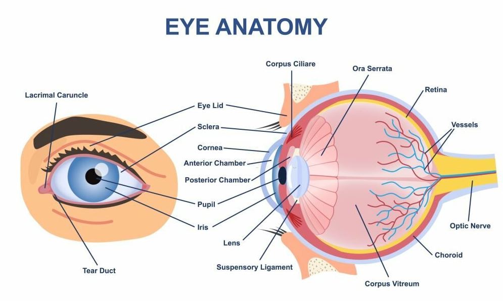
A knowledge of the anatomy of the eye is important to the understanding of eye diseases. A brief introductory outline of the structure and function of the eyeball and the tissues and structures around it is given below.
The eyeball consists of three tunics (coats) or ‘parts’.
Tunica fibrosa
The fibrous coat is the tough outer coat which is transparent anteriorly (the cornea) and opaque posteriorly (the sclera). The two meet at a junction called the limbus. The cornea helps in the refraction of light into the eye (about 2/3rd). The transparent nature of it is a result of the regular arrangement of collagen fibres in the stroma (the middle section of the cornea). The cornea is significantly avascular (meaning, no blood vessels) hence it is nourished by the tears anteriorly and the aqueous posteriorly. Also, the sclera which is the toughest part of the eye gives the shape of the eyeball and it is opaque because of the irregular arrangement of its collagen fibres. It is vascularized, meaning it has blood vessels running through it.
Tunica vasculosa
The vascular coat is rich in blood vessels and lies between the two coats. It consists of the more posteriorly choroid (nourishes the retina), the intermediate ciliary body (ciliary muscle – accommodation and ciliary processes – aqueous humour production) and the iris which has a central aperture called the pupil to regulate the amount of light that enters the eye. The iris gives the colour of the eye – brown, grey, amba, blue, green, hazel. Suspensory ligaments (zonules) arising from the ciliary processes hold the crystalline lens in place behind the iris.
The iris attaches itself to the cornea forming the iridocorneal angle lined by a meshwork of cells and collagen beams (trabecular meshwork). The aqueous humour drains here into the canal of Schlemm into the venous system. From the posterior side of the cornea to the anterior part of the iris is the anterior chamber and from the iris to the lens is the the posterior chamber. Aqueous humor fills both of these chambers. Between the lens and the retina lies the vitreous body.
Tunica nervosa
The inner layer is the retina which lines the back two-thirds of the eye. It consists of the retinal pigmented epithelium (RPE) and the neuroretina. The RPE consists of a monolayer of pigmented cells situated between the neuroretina and the choroid.
The RPE:
- separates the fenestrated endothelium of the choriocapillaries from the neuroretina (outer blood-retina barrier). The inner BRB is mainly constituted by endothelial cells (through tight junctions) of blood vessels and neighbouring neuroretina cells.
- transport nutrients, ions and water.
- isomerization of all-trans-retinal into 11-cis-retinal, which is a key element of the visual cycle.
- phagocytosis of shed photoreceptor membranes
- secretion of various essential factors for the structural integrity of the retina.
One key aspect of the anatomy of the eye is the neuroretina which consists of the other nine segments of the retina. The main connection is from photoreceptor cells (rods and cones) to bipolar cells to ganglion cells. The photoreceptor cells capture light and convert it into electrical signals, a process known as phototransduction. The bipolar cells provide the main pathways from photoreceptors to ganglion cells. The axons of the ganglion cell form the nerve fibres of the retina which exit the eye through the optic disc as the optic nerve. These transmit the electrical signals from the bipolar cells to the brain through the visual pathway. The macula (with a central pit called the fovea) is for detailed central vision. The peripheral retina is for dim light (or night) vision.
The two optic nerves decussate at the optic chiasm where both nasal nerve fibres cross to the contralateral optic tract. Temporal fibres do not cross hence each optic tract views the contralateral visual field. The optic tract ends at the lateral geniculate body (LGB). These send optic radiations through the brain to the striate cortices and extrastriate areas for decoding the electrical visual signals.
The Extraocular Muscles
The globe is moved by six extraocular muscles: four recti muscles and two oblique muscles, that is, the superior rectus, inferior rectus, medial rectus, lateral rectus, superior oblique and inferior oblique muscles. The four recti muscles have their origin in the common tendinous ring (annulus of Zinn) at the apex of the orbit which is connected to the dural sheath of the optic nerve. All four of the recti muscles project forward within a sheath derived from Tenon’s capsule and attach to the globe. On reaching the globe, each extraocular muscle inserts at different points behind the corneal limbus forming a spiral called the spiral of Tillaux.
The superior oblique originates external to the CTR, from the body of the sphenoid bone. Its origin is superonasal to the CTR and the optic nerve. The body of the muscle then passes forward and medially to bend around a connective-tissue pulley known as the trochlea. It enters the Tenon’s capsule before passing underneath the superior rectus and attaches to the globe. Of the six EOMs, the inferior oblique is the only muscle to originate from the front of the orbit. It arises from the orbital floor lateral to the nasolacrimal canal and just within the orbital rim. It then moves below the inferior rectus to curve upwards around the globe. Overlying the attachment is the lateral rectus.
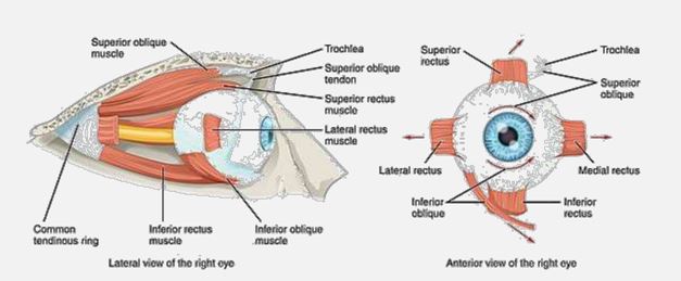
| EOM | Innervation (CN) | Function |
| Superior rectus | CNIII | Elevate, intort, adduct |
| Inferior rectus | CNIII | Depress, extort, adduct |
| Medial rectus | CNIII | Adduct |
| Lateral rectus | CNVI | Abduct |
| Superior oblique | CNIV | Intort, depress, abduct |
| Inferior oblique | CNIII | Extort, elevate, abduct |
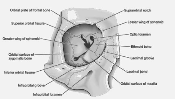
The Bony Orbit
The eye lies within the bony orbit (Fig. 1.2). The orbit is a bony pyramid with four walls: a roof, lateral wall, floor and medial wall. The base of the pyramid is the entrance of the orbit which is roughly rectangular. At its posterior apex is the optic canal which transmits the optic nerve to the brain. The superior and inferior orbital fissures allow the passage of blood vessels and cranial nerves which supplyorbital structures. On the anterior medial wall lies a fossa for the lacrimal sac. The lacrimal gland lies anteriorly in the superolateral aspect of the orbit. The bony orbit also serves as a point of attachment of the six extraocular muscles. The bony orbit is covered by the periosteum which is continuous with the optic nerve as the dura mater. The rest of the orbital cavity is filled with fat (orbital fat).
Walls of the orbit:
- Roof (superior): orbital part of the frontal bone, lesser wing of the sphenoid bone.
- Medial: orbital plate of the ethmoid bone, lacrimal bone, frontal process of the maxilla, lesser wing of the sphenoid bone.
- Floor (inferior): the orbital surface of the maxilla, zygomatic bone, and palatine bone.
- Lateral: zygomatic bone, sphenoid bone.
The Adnexae
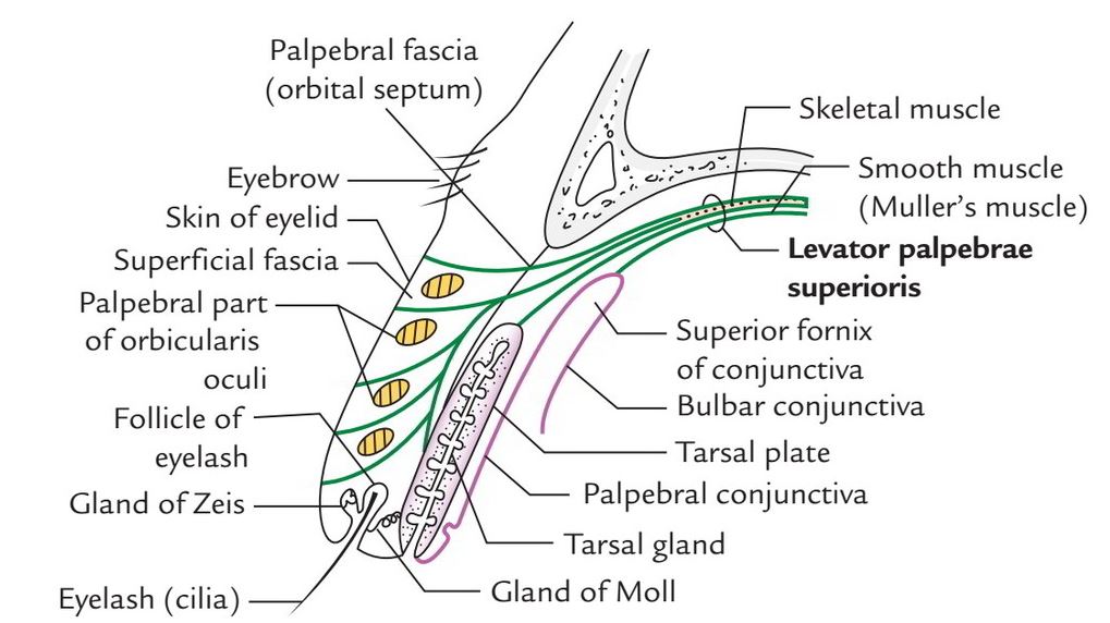
The adnexae consist of the eyebrow, the eyelid, and the lacrimal system. Eyebrows are two arched ridges of the supraorbital margins of the frontal bone. Numerous hair (eyebrows) project obliquely from the surface of the skin. They protect the eyeball from sweat, dust and other foreign bodies.
The eyelids are two movable folds of tissue situated above and below the front of the eye. There are short curved hairs, and the eyelashes situated on their free edges.
The eyelid consists of:
- A thin covering of skin
- Three muscles—the orbicularis oculi, levator palpebrae superioris and Müller’s muscles
- A sheet of dense connective tissue, the tarsal plate
- A lining of the conjunctiva.
The conjunctiva, a thin, translucent mucous membrane, extends from the limbus through the fornices to the lid margin and can be divided into three parts:
- Bulbar conjunctiva
- Palpebral conjunctiva
- Forniceal conjunctiva
The conjunctiva ensures a smooth movement of the eyelid over the globe as well as forms a barrier which prevents entry into the orbit from outside.
The Lacrimal apparatus consists of:
- Lacrimal gland and its ducts
- Accessory lacrimal glands
- Lacrimal canaliculi
- Lacrimal sac
- Nasolacrimal duct
The tears are secreted by the lacrimal gland and accessory lacrimal glands. They drain into the conjunctival sac and then pass into the lacrimal sac via the canaliculi, moves on to the nasolacrimal duct and finally into the nasal cavity.
Blood and Nerve Supply to the Orbit
The arterial supply of the eye is by the ophthalmic artery, a branch of the internal carotid artery. This branches into the short (about 20 in number) and long ciliary (2 in number) arteries and the central retinal artery. The short ciliary veins, anterior ciliary veins, 4 vortex veins, and the central retinal vein actively perform venous drainage. These eventually empty into the cavernous sinus.
The eye is supplied by three types of nerves, namely motor, sensory and autonomic.
1. The Motor Nerves
- The third cranial nerve (oculomotor) –- it supplies the superior, inferior, medial recti and the inferior oblique muscles. It also supplies the levator palpebral superioris.
- The 4th cranial nerve (trochlear)—it supplies the superior oblique muscle.
- The 6th cranial nerve (abducens)—it supplies the lateral rectus muscle.
- The 7th cranial nerve (facial)—it supplies the orbicularis oculi muscle.
2. The Sensory Nerve
- The 5th cranial nerve (trigeminal)—the ophthalmic division supplies the whole eye.
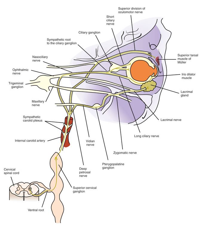
3. The Autonomic Nerves
a. The sympathetic nerve supply is through the cervical sympathetic fibres to:
- Iris—dilator pupillae muscle
- Müller’s muscle in the lids
- Lacrimal gland.
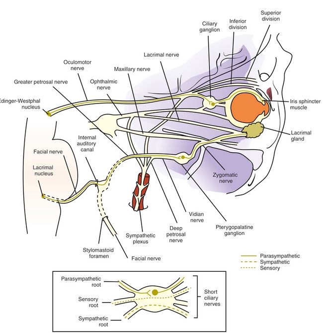
b. The parasympathetic nerve supply originates from the nuclei in the midbrain:
- Iris—sphincter pupillae muscle (oculomotor)
- Ciliary body (oculomotor)
- Lacrimal gland (facial nerve).
This is an introduction to the anatomy of the eye.
By the way, The expression ‘the eyes are the windows to the soul’ is metaphorical, conveying the idea that a person’s eyes can unveil their genuine emotions, feelings, and intentions. It suggests that gazing into someone’s eyes can give you a glimpse into their innermost thoughts and emotions.”